Describe the Lens of the Dissected Eye
What is the function of the dark pigment in the choroid coat. Experts are tested by Chegg as specialists in their subject area.

Sheep Eye Flashcards Quizlet
Describe the function of three areas of the brain you choose which areas you wish to discuss.
. A anterior portion 10x. The cloudy condition called cataract prevents or reduces the amount of light reaching the retina. Cerebrum- vision hearing emotions movement.
Describe the vitreous humor of the dissected eye. The vitreous humor is a clear gelatinous material. Waste products are removed through.
Lens It is clear and flexible that makes an image on the retina and since its flexible it can change shape to focus on objects near or far. The normal lens is convex shaped and somewhat elastic. B posterior portion 53x.
It felt hard like a marble but it was not shaped exactly spherical like a marble. Describe the iris and explain its function. What is the lens of the eye.
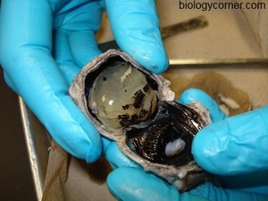
Up to 24 cash back The pupil is the dark circle thats in the center of the iris and it lets light into the inner eye. Using a sharp scalpel cut through the sclera around the middle of the eye so that one half will have the anterior features of the eye the cornea lens iris and ciliary body and the other half will contain the posterior features most noticeably where the optic nerve is attached to the eyeThe inside of the eye cavity is filled with liquid. It is located behind the iris and in front of the vitreous body.
Ask the students what they see when they hold up the lens and look through it They see. Where do you find aqueous humor in the dissected eye. Order of light passing through eye.
Where was the aqueous humor in the dissected eye. Cataract can be treated by removing the lens and replacing it with a stiff artificial one. Describe the vitreous humor of the dissected eye.
Preservatives make the lens hard and opaque but in living organisms the lens is clear and fl exible. And when there is low light the iris opens up the pupil to let in more light. Lens in anatomy a nearly transparent biconvex structure suspended behind the iris of the eye the sole function of which is to focus light rays onto the retina.
The eyes lens uses the light that comes into the eye to make an image a picture made of light. Virtual Eye Dissection and Eye Anatomy Tools. The retina is located ahead of the point where the lens and cornea produce a focused image.
Describe the tissue of the lens you felt during the dissection of the cow eye. The lens is a transparent structure in the eye that along with the cornea helps to refract and focus light. Middle of the eye diagram behind the lens and then show the image right-side-up on the retina the way it is.
Image of dissected eye with the lens removed with your name and access code handwritten clearly in the background. A step-by-step hints and tips a cow eye primer and a glossary of terms. Describe the lens of the dissected eye sheep eye This problem has been solved.
Describe the vitreous humor. Use your finger to push the retina around. What is the function of the dark pigment in the choroid coat.
To help us focus on closer objects is gets fatter to refract more light and vice versa for focusing on. The cells of the retina react to the light that falls on them and send messages to the brain. The lens of the eye also called the crystalline lens is an important part of the eyes anatomy that allows the eye to focus on objects at varying distances.
Image of dissected eye with the lens removed with your name and access code clearly visible in the background. In its natural state the lens looks like an elongated sphere a shape known as ellipsoid that resembles a deflated ball. See the answer See the answer See the answer done loading.
It is clear so that light can pass through it to the retina. That image lands on the retina. Dry the eye with paper toweling.
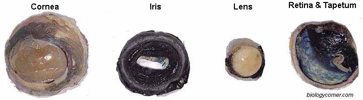
Learn how to dissect a cows eye in your classroom. The lens focuses light onto the retina. The colored part of the eye which helps regulate the amount of light entering the eye.
A diverging lens placed in front of the eye causes the rays entering the eye to spread out so that the cornea and the lens focus the light correctly and form a clear image on the retina. We review their content and use your. Sheep eye dissecting pan surgeons gloves scissors single edge razor blade probe forceps paper towels and a notebook and pencil for recording information about the eye as it is dissected Step 1.
Describe the lens of the dissected eye. Describe the vitreous humor of the dissected eye. Brain Mapping Post-Lab Questions 1.
Describe the lens of the dissected eye. List the two functions of the lens. It felt hard like a marble but it was not shaped exactly spherical like a marble.
When there is bright light the iris closes the pupil to let in less light. The retina is made of cells that can detect light. O The lens of the cows eye feels soft on the outside and hard in the middle.
How do you compare the shape of the pupil in the dissected eye with your pupil. Figure 3510 Sections of the eye. The lens is made up of unusual elongated cells that have no blood supply but obtain nutrients from the surrounding fluids mainly the aqueous humour that bathes the front of the lens.
Who are the experts. Lens Focuses light rays onto the retina. Describe the lens of the dissected eye.
That image lands on. Cows Eye Dissection Lab Page 2. Describe the lens of the dissected eye sheep eye Expert Answer.
Normal condition the lens is transparent except when as a condition of aging the lens turns cloudy. Describe the function of three areas of the brain you choose which areas you wish to discuss. Wash the sheep eye in running water to remove the preservative fluid.
The lens is transparent and can be replaced if necessary. The Cow Eye Dissection Lab. The eyes lens uses the light that comes into the eye to make an image a picture made of light.
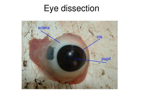
Describe the iris and explain its function. Cows Eye Dissection - Eye diagram. The eye of a person who is farsighted is slightly shorter than normal.
O The lens is a transparent sphere that can change its shape to focus the light entering the eye. Examine the front of the eye and locate the eye-lid cornea sclera. Opportunities to describe how light passes through the parts of the eye and is received by the brain.
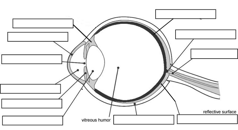
Cornea - aqueous humor - pupil - lens - vitreous humor - retina. The retina is made of cells that can detect light. Why must the vitreous humor be clear.
Cow Eye Dissection

Ha 26p Eye Dissection Lab Document Pdf Name Block Date 2114120 Juliette 5 Hill Honors Anatomy Physiology Eye Dissection Lab Document 1 Label The Course Hero

Sheep Eye Dissection Photos Flashcards Quizlet

Zebrafish Lens Dissection A Eyes Are Dissected From Anesthetized Download Scientific Diagram
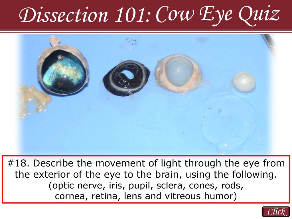
Cow Eye Quiz Dissection 101 Click Ppt Download

Cow Eye Dissection Nandini Soni
Cow Eye Dissection
9 Lenses And The Eye Wk6 Mrs Morritt Science
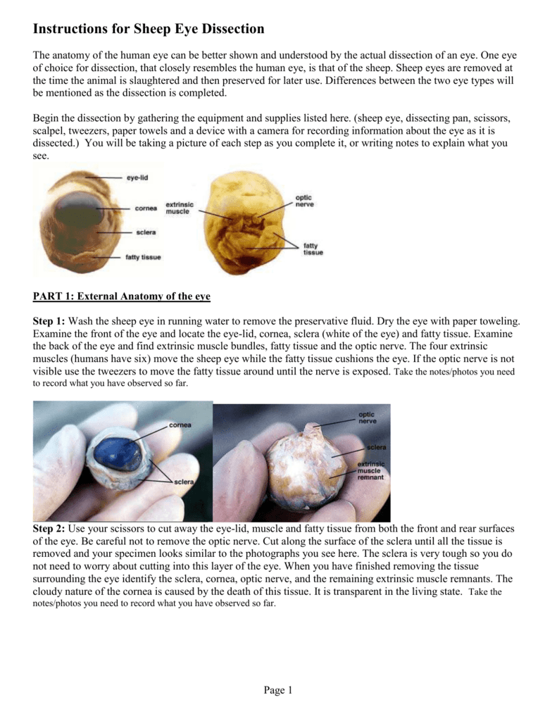
Instructions For Sheep Eye Dissection
Cow Eye Dissection Welcome To Cow Eye Dissection This Powerpoint Was Created To Be Downloaded And Used With Your Students Please Feel Free To Edit Ppt Download

Cow Eye Dissection Perkins Elearning
2

Sheep Eye Lab Directions

Cow S Eye Dissection Step 8
Cow Eye Dissection
2

Cow Eye Dissection Nandini Soni

Frog Anatomy Review Pig Dissection Dissection Animal Science

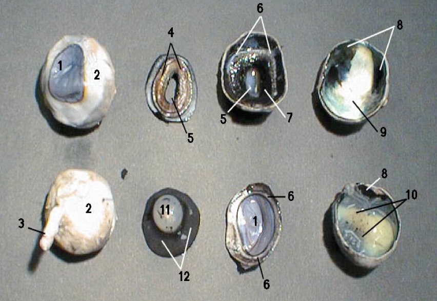
Lab 12 Sheep Eye Dissection Diagram Quizlet


Comments
Post a Comment